Medial canthal entropion in pugs
What is the medial canthal entropion of the eyelid?
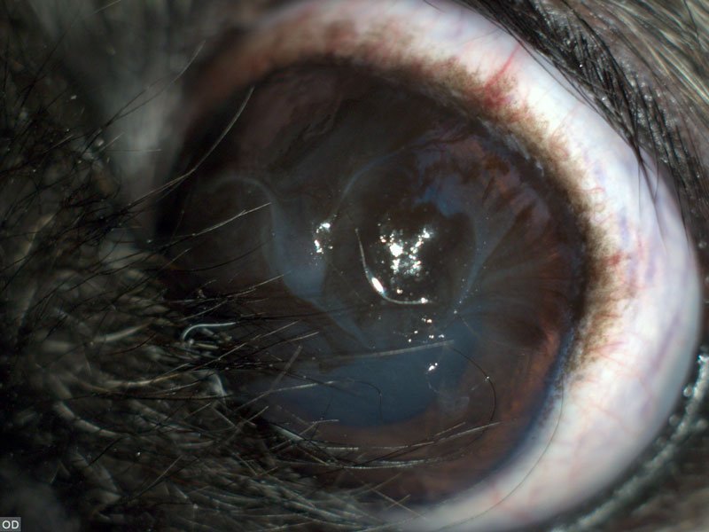
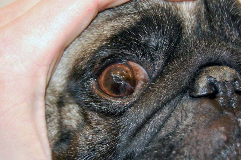
Medial entropion is a phenomenon in which the eyelid at the inner corner of the eye (corner adjacent to the nasal region) is rotated inward. This results in direct contact of the hair of the
rotated eyelid and corneal surface, which further results in corneal irritation, inflammation and/or ulceration. This is a common condition in brachiocephalic breeds of dogs, especially in Pugs, Pekingese, Shih Tzu, but also in brachiocephalic breeds of cats (Persian cat).
Brachiocephalic breeds have a number of other eye problems which are contributing to medial canthal entropion syndrome, and these are:
- Long eyelids
These breeds have too long eyelids that allow excessive exposure of the eye to external influences.
- Thin eyelid margin
The eyelid margin is the boundary between the haired skin on the surface of the eyelid and conjunctiva on the inner surface of the eyelid. In brachiocephalic breeds, the eyelid margin is too thin, and in some dogs non-existent, which allows rotation of the eyelid toward the surface of the eye.
- Shallow orbit
Brachiocephalic dogs have a shallow orbit, which provides less sufficient protection to the eye than a deeper orbit in other breeds of dogs. The shallow orbit results in “bulging” appearance of the eyes and lagophthalmos (inability to completely close the eyelids). This is why the eyelids in brachiocephalic breeds fail to fully protect the eye surface and allow the exposure of cornea to the external adverse influences.
- Trichiasis
The medial caruncle (also called lacrimal caruncle) is a small nodule located at the inner corner of the eye (medial cantus of the eye). The medial caruncle is composed of skin (which includes hair), lacrimal glands, and other tissues. In brachiocephalic breeds, medical caruncle often contains excessive hair, which rubs the surface of the eye (trichiasis) resulting in corneal irritation, inflammation and/or ulceration.
- Reduced innervation of the cornea
The innervation of the cornea is reduced in brachiocephalic breeds, which is the reason why these dogs do not sense the pain and express discomfort that other breeds of dogs would clearly express. Therefore, the owners notice clinical symptoms when the disease has progressed to a later stage, and seek help of a veterinary ophthalmologist only when they notice “spots” on the cornea.
- Dry eye disease
In brachycephalic breeds, qualitative or quantitative tear film deficiency is a common issue which may result in dry eye disease.
- Excessive tearing (epiphora)
The anatomic structure of the head, especially the orbit in brachiocephalic breeds, can lead to anatomical changes in the nasolacrimal duct, making it less passable for the tears. The opening of the nasolacrimal duct is additionally narrowed by medial entropion. In situations like this, the tears will accumulate at the corner of the eye, and instead of draining into the nasal or oral cavity, the tears will drain down the face diminishing the protective function to the cornea and resulting in dermatitis.

What are the consequences of all the above-mentioned conditions?
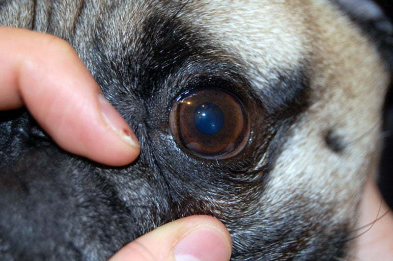
Over time, the irritation of the cornea results in pigmentary keratitis which clinically appears as rough and dark cornea. The pigment initially accumulates at the medial side of the cornea, and over time gradually affects the larger corneal surface. Therefore, the dogs gradually become sluggish, reluctant to run, and uninterested in everyday activities, as they can only recognize shadows and feel insecure. The owners can also notice a discharge or formation of the thick crust around the eyes, especially in the morning, and the dog appears the have difficulties opening the eyelids. In some pugs, these changes are noticed as early as one or two years of age.
What can be done to prevent pigmentary keratitis?
The surgical correction of the medial entropion can be done in order to eliminate all the changes that occur in brachiocephalic ocular syndrome.
- Entropion surgery
The surgery will correct the entropion, and the hair will be removed from the medial caruncle
- Shortening of the eyelids and correction of the eyelid
The eyelids will be shortened eyelid margin will be corrected. Shortening the eyelids corrects the problem of too long eyelids, and eyelids regain the role of eye protection.
- Reduction of palpebral opening
By reducing the palpebral opening (the opening that covers the eyelids), the probability of prolapse of the eye, which often occurs as a consequence of trauma in brachiocephalic breeds, is decreased.
- Nasolacrimal duct opening/widening
The opening of the nasolacrimal canals is released, which facilitates the drainage of tears from the eye into its natural drains and better lubrication of the cornea with the tear film.

How can the owner recognize this problem and timely react?
If you are the owner of a pug or other brachiocephalic breed, consider that your dog certainly has medial entropy or ocular syndrome of brachiocephalic breeds. You can see it easily. Take your pet and evaluate its eyes with a bright source of light. Start by carefully observing the edge of the lower eyelid along the nasal fold. You will see that the eyelid hairs rub the cornea and that the eyelid margin of the lower eyelid is very thin until it almost completely disappears where the lower eyelid joins the upper eyelid. After that, evaluate the cornea adjacent to the eyelid. You will see the dark spots on the corneal surface. Compare it to the cornea along the lateral edge of the eyelid (the edge facing the ear). Sometimes the entire cornea will be darkly pigmented, so there will be no difference, but then you will realize that you cannot see the iris (the colored part of the eye). In such dogs, the pupil and iris are not visible, and everything is equally dark due to the severe pigmentation of the cornea. The sooner you notice a problem; the sooner you will be able to resolve it. The success of treatment primarily depends on the stage of the pathological processes. If the medial entropion of the eyelid is corrected at an early age, then there is a high probability that such a dog will not have consequences for the rest of its life. If the medial entropion of the eyelids has already caused serious corneal lesions, the surgery will prevent irritation of the cornea by the hair; however, lifelong use of topical eye medication in the form of drops will be necessary. If the vision is significantly impaired, surgery is needed to correct the medial entropion and remove corneal tissue affected by the pigmentary keratitis; however, lifelong therapy with local medication will be needed.
When to perform surgery to correct the medial entropion?
The optimal age for performing surgery in a pug with medial canthal syndrome is at 7-10 months of age. This is because this is the age in which first corneal lesions typically appear.
Course of the recovery from the surgery?
After the surgery, the dog will experience no pain; however, wearing the protective collar and administering appropriate medication will be needed. After 14 days, the dog will not require any special care.


